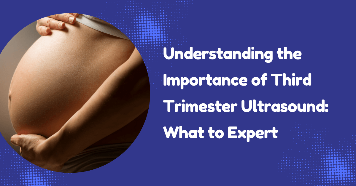
The third trimester, spanning from 28 to 40 weeks of pregnancy, marks a vital and transformative period for both mother and baby. During these weeks, your baby experiences significant growth and development as they prepare for their entrance into the world.
Growth Scans, Fetal Wellbeing & Late Pregnancy Monitoring (28–40 Weeks)
A third-trimester ultrasound, often referred to as a growth scan, plays an essential role in this stage by offering a comprehensive view of your baby’s size, position within the womb, and overall health. This detailed imaging not only provides reassurance to expectant parents but also helps healthcare providers monitor any important changes, ensuring that everything is progressing smoothly as the due date approaches.
If you’re in Leeds or West Yorkshire, we offer expert
private pregnancy ultrasound scans in Leeds to monitor your baby’s development during the final stage of pregnancy.
The third trimester (28–40 weeks) is a critical phase of pregnancy where your baby undergoes rapid growth and preparation for birth. A third-trimester ultrasound (growth scan) provides detailed insights into your baby’s size, position, and overall well-being.
This scan is essential for ensuring that your baby is developing appropriately and for identifying any potential concerns early. It is often recommended for reassurance, especially if there are clinical concerns or previous pregnancy complications.
👉 Combine your scan with pregnancy blood tests in Leeds
for a complete maternal and fetal assessment.
Why Is a Third Trimester Ultrasound Important?
Each of these parameters provides important insights into the fetal growth pattern and overall health, helping healthcare professionals assess whether the fetus is developing appropriately for its gestational age. Additionally, the scan allows for observation of other factors such as amniotic fluid levels and the position of the placenta, further contributing to a comprehensive understanding of the pregnancy.
- Assess fetal growth and weight
- Monitor amniotic fluid levels
- Evaluate placental position and function
- Check fetal position (head-down, breech)
- Identify potential complications before delivery
This scan provides reassurance and helps healthcare professionals plan a safe delivery.
Monitoring Baby’s Growth & Development
During the ultrasound scan, a variety of crucial measurements are taken to evaluate the growth and development of the fetus. These measurements include the biparietal diameter (the width of the head), the head circumference (the distance around the head), the abdominal circumference (the measurement around the belly), and the femur length (the length of the thigh bone).
- Head circumference (HC)
- Abdominal circumference (AC)
- Femur length (FL)
These measurements are used to estimate your baby’s weight and ensure they are growing appropriately for gestational age.
👉 Growth scans are often combined with advanced pregnancy ultrasound packages
Amniotic Fluid Assessment
Amniotic fluid plays a vital role in protecting your baby and supporting development. Ultrasound is used to assess fluid levels:
- Low levels (oligohydramnios) may indicate placental issues
- High levels (polyhydramnios) may require further monitoring
Placental Function & Position
The placenta supplies oxygen and nutrients to your baby. During the third-trimester scan, we assess:
- Placental location (e.g. low-lying placenta)
- Blood flow and function
- Signs of placental insufficiency
Fetal Wellbeing Assessment
The ultrasound allows real-time evaluation of:
- Fetal movements
- Breathing patterns
- Heart activity
- Overall wellbeing
This provides reassurance and helps detect any concerns early.
When Should You Have a Third Trimester Scan?
Most scans are performed between 28 and 40 weeks, commonly around 32 weeks.
You may need additional scans if:
- Reduced fetal movements
- Previous complications
- Growth concerns
- High-risk pregnancy
What to Expect During the Scan
The procedure is safe, painless, and similar to earlier scans:
- Gel is applied to your abdomen
- A probe (transducer) captures images
- Scan takes approximately 15–30 minutes
Optional 3D/4D scans may also be available for enhanced visualisation.
Private Pregnancy Scans in Leeds
- 👉 Third Trimester Growth Scans
- 👉 Pregnancy Blood Tests
- 👉 Same-day appointments available
Serving Leeds, Bradford, Wakefield & West Yorkshire.
Third Trimester Ultrasound FAQs
- When should I have a third-trimester scan?
Usually between 28–32 weeks, but can be done up to 40 weeks. - Is the scan safe?
Yes, ultrasound uses sound waves and is completely safe. - Can it detect problems?
Yes, it helps identify growth issues, fluid abnormalities, and placental problems. - Do I need a scan if everything is normal?
Many parents choose it for reassurance, even in low-risk pregnancies.
Serving Leeds & West Yorkshire
We provide private pregnancy ultrasound scans in Leeds, easily accessible for patients across West Yorkshire.